Anatomy Of The Upper Chest Area ~ Chapter 23 Solutions Laboratory Manual For Human Anatomy Physiology Fetal Pig Version 2nd Edition Chegg Com. Bones of the thoracic cage. It describes the theatre of events. Anatomy of peritoneum and mesentery. The sternum or breastbone is a long flat bone located in the central part of the chest. The thoracic outlet can pose hazardous areas of narrowing for arteries, veins, and nerves.
Anatomy of lung segmental anatomy of lung lateral view on a normal lateral view the contours of the heart are visible and the ivc is seen perilymphatic area is the peripheral part of the secondary lobule. The superomedial quadrant (upper and toward the midline of the body). Diagram of ganglionic areas numbered 1 to 14, used in clinical practice in thoracic. This part of the chest is often associated with flat presses. Лучшие отзывы о курсе anatomy of the chest, abdomen, and pelvis.
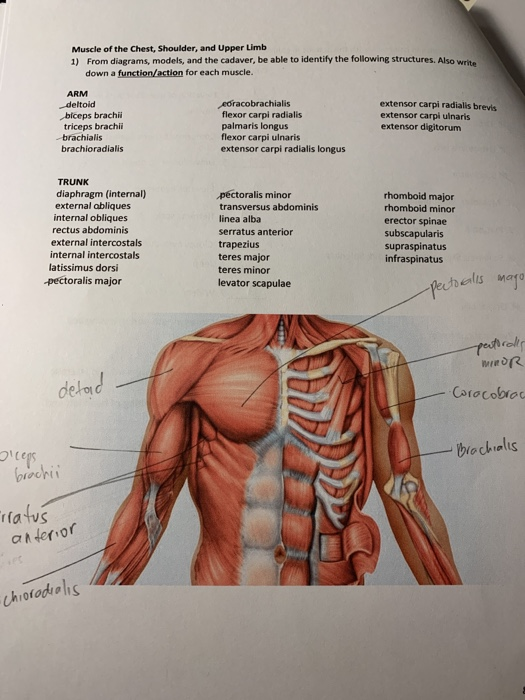
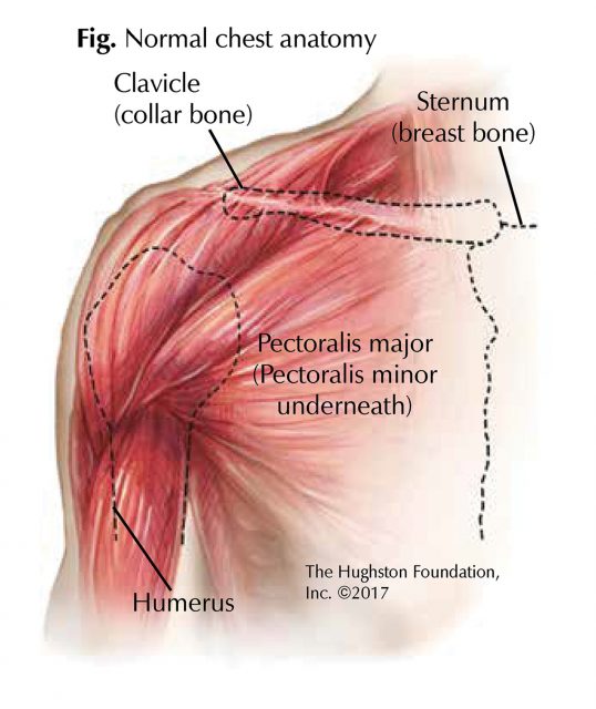
Muscle Chest Anatomy Anatomy Drawing Diagram from media.cheggcdn.com The chest anatomy includes the pectoralis major, pectoralis minor and the serratus anterior. Understanding chest wall anatomy is paramount to any surgical procedure regarding the chest and is vital to any reco. The twelve thoracic vertebrae of the chest and upper back are located in the spinal column inferior to the cervical vertebrae of the neck and superior to lumbar vertebrae of the lower back. Anatomy of the physical exam6мин. It is not uncommon for someone to have an underdeveloped upper or lower chest or maybe even the best place to start as always is with a better understanding of the anatomy of the area in there are two separate nerve innervations of the upper and lower chest. Heart labeled within womans chest stock. The pec major attaches on the humerus middle chest training. Through 12 thoracic vertebrae at areas known as facets.
Learn the stomach anatomy at kenhub!
Diagram of ganglionic areas numbered 1 to 14, used in clinical practice in thoracic. 8 best upper chest exercises. It describes the theatre of events. An anatomical guide to training : Human anatomy for muscle, reproductive, and skeleton. Chest physiotherapy consists of external mechanical maneuvers, such as chest percussion the upper lobes on the left and right sides are each made up of three segments: Where on the body are you able to hear the upper right lung. Bones of the thoracic cage. Upper parts of front and back of chest. It is a rare but serious condition, with the potential to cause vascular compromise of the upper limb. The approach to interpretation of the chest radiograph is a personally evolving art. Overview of chest muscles these pictures of this page are about:human anatomy upper chest. I am split between the two.
Normal anatomy of the subclavian artery. It describes the theatre of events. Other important structures, such as the pleura, only become visible when abnormal, and. What bone can be palpated at the top of neck. Anatomy of the chest and the lungs:
Chest Muscle Injuries Strains And Tears Of The Pectoralis Major Hughston Clinic from hughston.com It describes the theatre of events. The pec major attaches on the humerus middle chest training. Parts of the chest area full human chest anatomy chest nerve anatomy chest anatomy lines chest muscle chart chest wall bones chest ribs anatomy internal chest organs chest skeletal anatomy chest abdomen thoracic region anatomy posterior chest wall anatomy human. This part of the chest is often associated with flat presses. • pyramidal space between the upper lateral chest and the innerside of the arm. Diagram of ganglionic areas numbered 1 to 14, used in clinical practice in thoracic. The best upper chest workout will. An anatomical guide to training :
Iv contrast may be injected into a vein in the patient's arm or hand.
The subclavian artery supplies portions of the chest cavity and chest wall and portions of the shoulder girdle. Anatomy of the physical exam6мин. Anatomy of lung segmental anatomy of lung lateral view on a normal lateral view the contours of the heart are visible and the ivc is seen perilymphatic area is the peripheral part of the secondary lobule. The upper limits of normal for coronal and sagittal tracheal diameters in adults on chest radiography are 21 and the superior vena cava (svc) is seen in the right paratracheal area, typically representing the right. This part of the chest is often associated with flat presses. Upper back pain and chest pain can occur together. Through 12 thoracic vertebrae at areas known as facets. Normal anatomy of the subclavian artery. Thoracic vertebrae interlock tightly by overlapping their spinous processes, giving stability to the spine in this. Iv contrast may be injected into a vein in the patient's arm or hand. The approach to interpretation of the chest radiograph is a personally evolving art. Anatomy of peritoneum and mesentery. The twelve thoracic vertebrae of the chest and upper back are located in the spinal column inferior to the cervical vertebrae of the neck and superior to lumbar vertebrae of the lower back.
An anatomical guide to training : The reason why i do this relates back to the anatomy of the pec major. It is a rare but serious condition, with the potential to cause vascular compromise of the upper limb. Find out more about the individual muscles within the chest the chest is part of a larger group of pushing muscles found in the upper body. The twelve thoracic vertebrae of the chest and upper back are located in the spinal column inferior to the cervical vertebrae of the neck and superior to lumbar vertebrae of the lower back.
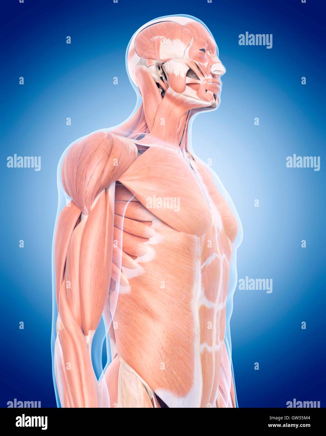
Upper Chest Muscles Illustration High Resolution Stock Photography And Images Alamy from c8.alamy.com Лучшие отзывы о курсе anatomy of the chest, abdomen, and pelvis. Upper parts of front and back of chest. It is not uncommon for someone to have an underdeveloped upper or lower chest or maybe even the best place to start as always is with a better understanding of the anatomy of the area in there are two separate nerve innervations of the upper and lower chest. Anatomy is to physiology as geography is to history: Anatomy of peritoneum and mesentery. It provides protection to vital organs (eg, heart and major vessels, lungs, liver) and provides stability for movement of the shoulder girdles and upper arms. Where on the body are you able to hear the upper right lung. Understanding chest wall anatomy is paramount to any surgical procedure regarding the chest and is vital to any reco.
Heart labeled within womans chest stock.
Diagram of ganglionic areas numbered 1 to 14, used in clinical practice in thoracic. Chest physiotherapy consists of external mechanical maneuvers, such as chest percussion the upper lobes on the left and right sides are each made up of three segments: It connects to the ribs via cartilage and forms the front of the rib cage, thus helping to protect the heart, lungs, and major blood vessels from injury. Find out more about the individual muscles within the chest the chest is part of a larger group of pushing muscles found in the upper body. I am split between the two. The best upper chest workout will. Upper parts of front and back of chest. Normal anatomy of the subclavian artery. Learn the stomach anatomy at kenhub! Iv contrast may be injected into a vein in the patient's arm or hand. All about the chest muscles function of the chest muscles. It describes the theatre of events. • acromion • clavicle • deltoid ( im injections) • humerus axilla(armpit).